
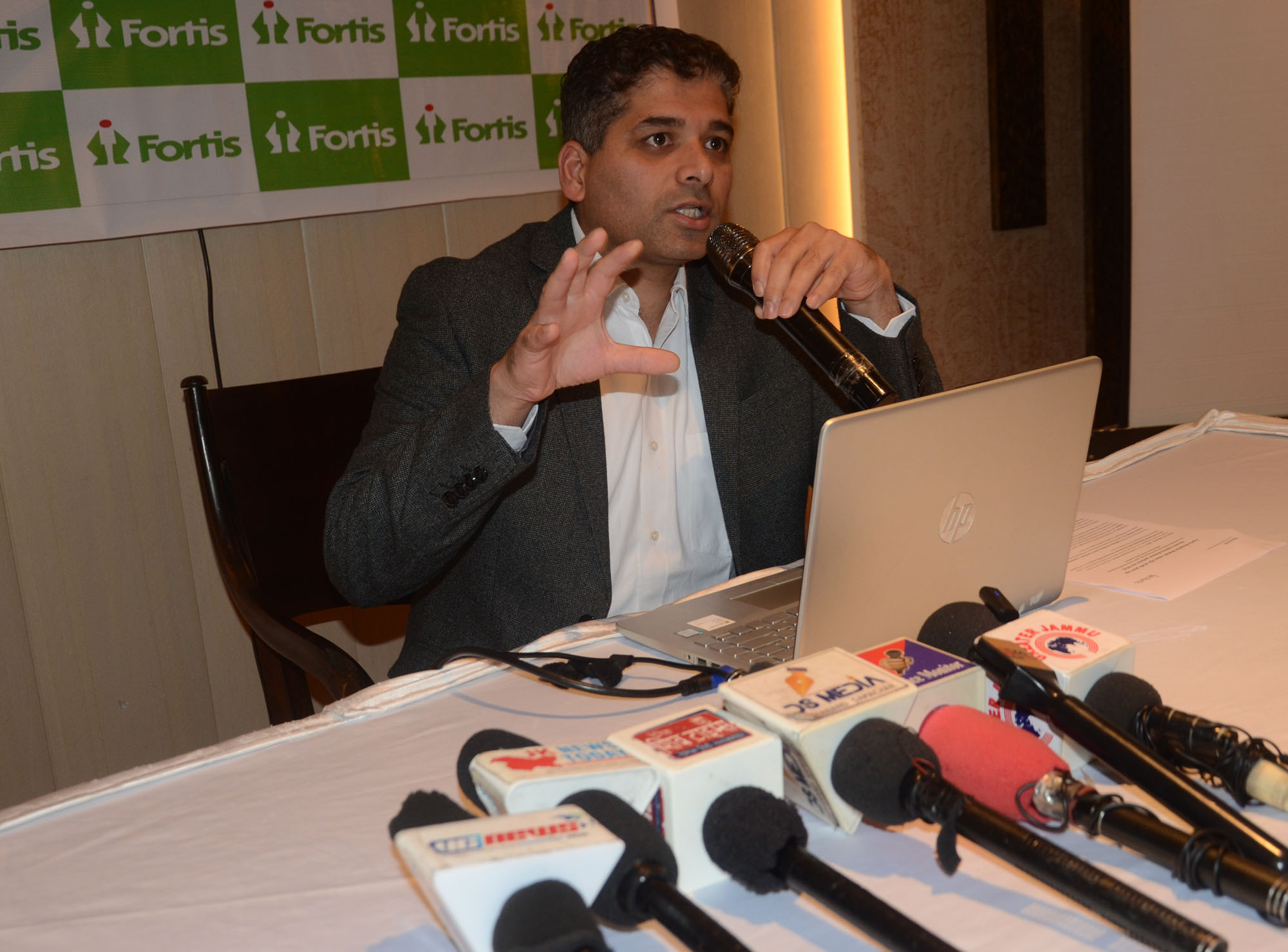
JAMMU, Sep 25: Doctors at Fortis Hospital, Mohali, successfully performed a complex coronary angioplasty on an 80-year-old man suffering from chronic kidney disease (CKD), using ultra-low contrast angioplasty guided by Intravascular Ultrasound (IVUS).
The technique is designed to minimise the amount of contrast dye used during coronary procedures, an important consideration in patients with impaired kidney function who may be at risk of contrast-associated kidney injury.
The procedure was performed by Dr Sudhanshu Budakoty, Senior Consultant, Cardiology, Fortis Hospital, Mohali. According to the hospital, the patient was discharged in stable condition two days after the procedure.
The patient had been living with chronic kidney disease for around five years and presented with high creatinine levels, severe chest pain, breathlessness and coronary artery disease. Given his kidney condition, the use of conventional angioplasty involving larger quantities of contrast dye was considered a significant concern.
The hospital said the combination of ultra-low contrast techniques and IVUS imaging enabled the medical team to assess and treat the coronary blockages while using only a minimal quantity of contrast dye.
Explaining the procedure, Dr Budakoty said the patient had significant coronary artery disease, while his CKD made conventional angioplasty more challenging. He said IVUS enabled the team to assess vessel dimensions, lesion characteristics and stent placement and expansion while keeping contrast use to a minimum.
“Advanced imaging modalities like IVUS and Optical Coherence Tomography (OCT) are routinely used at Fortis Mohali to treat high-risk cardiac patients,” Dr Budakoty said, adding that such technologies can assist doctors in managing complex coronary cases, including those involving patients with serious underlying conditions.
IVUS, or Intravascular Ultrasound, uses sound waves to generate images from inside the blood vessels. It allows cardiologists to assess the size and characteristics of coronary arteries and helps guide stent placement and expansion.
OCT, or Optical Coherence Tomography, is another intravascular imaging technique that uses light to produce high-resolution images of coronary arteries. It can assist doctors in evaluating lesions and guiding treatment.
The hospital stated that the availability of advanced imaging technologies and specialised cardiology services enables its team to manage complex interventional cardiology cases involving high-risk patients.
The case highlights the role of intravascular imaging and contrast-sparing approaches in selected patients with coronary artery disease and impaired kidney function, where reducing exposure to contrast dye may be clinically important.